
Fichier:MultiPhotonExcitation-Fig7-doi10.1186slash1475-925X-5-36.JPEG

Taille de cet aperçu : 665 × 599 pixels. Autres résolutions : 266 × 240 pixels | 533 × 480 pixels | 852 × 768 pixels | 1 136 × 1 024 pixels | 2 273 × 2 048 pixels | 2 992 × 2 696 pixels.
Fichier d’origine (2 992 × 2 696 pixels, taille du fichier : 1,93 Mio, type MIME : image/jpeg)
Ce fichier et sa description proviennent de Wikimedia Commons.
Description
| Description |
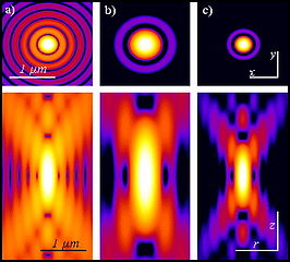
English: Original figure legend: Pointlike emitter optical response. From left to right: calculated x-y (above) and r-z (below) intensity distributions, in logarithmic scale, for a point like source imaged by means of wide-field, 2PE and confocal microscopy. Both 2PE and confocal shapes exhibit a better signal to noise ratio than widefield case. 2PE distribution is larger due the fact that a wavelength twice than in the wide-field and confocal case is responsible for the intensity distribution. Such intensity distributions are also known as point spread functions of the related microscopes. Optical conditions: excitation wavelengths are 488 nm and 900 nm for 1PE and 2PE, respectively; emission wavelength is 520 nm; numerical aperture is 1.3 for an oil immersion objective with oil refractive index value set at 1.515.
Deutsch: Original Abbildungslegende: Optische Antwort eines punktförmigen Emitters. Von links nach rechts: berechnete xy (oben) und rz (unten) Intensitätsverteilung in logarithmischer Skala für eine punktförmige Quelle aufgenommen in einem Weitfeld-, 2-Photonenanregungs- und Konfokal-Mikroskop. Sowohl die 2-Photonenanregungs, als auch die konfokalen Verteilungen zeigen ein besseres Signal-zu-Rausch-Verhältnis als im Fall des Weitfeldbildes. Das 2Photonenanregungsbild is weiter weil die benutzte Wellenlänge im Vergleich doppelt so groß ist. Diese Verteilungen sind auch als Punktspreizfunktionen ihrer Mikroskope bekannt. Optische Parameter: Anregungswellenlänger bei 488 nm für 1 Photonenanregung und 900 nm für 2 Photonenanregung; Emissionswellenlänge bei 520 nm und numerische Apertue 1.3 für ein Öl-Immersionsobjektiv mit einem angenommenen Brechungsindex für Öl bei 1.515. |
| Date | |
| Source |
Multi-photon excitation microscopy. BioMedical Engineering OnLine, 2006, 5:36. |
| Auteur | Alberto Diaspro, Paolo Bianchini, Giuseppe Vicidomini, Mario Faretta, Paola Ramoino and Cesare Usai |
| Autorisation (Réutilisation de ce fichier) |
Ce fichier est disponible selon les termes de la licence Creative Commons Attribution 2.0 Générique.
|
All images uploaded from this article about multi-photon and two-photon-microscopy:


Historique du fichier
Cliquer sur une date et heure pour voir le fichier tel qu'il était à ce moment-là.
| Date et heure | Vignette | Dimensions | Utilisateur | Commentaire | |
|---|---|---|---|---|---|
| actuel | 23 décembre 2008 à 20:57 | | 2 992 × 2 696 (1,93 Mio) | Dietzel65 | == Beschreibung == {{Information |Description={{en|1=Original figure legend: ''Pointlike emitter optical response. From left to right: calculated x-y (above) and r-z (below) intensity distributions, in logarithmic scale, for a point like source imaged by |
Utilisation du fichier
La page suivante utilise ce fichier :
Usage global du fichier
Les autres wikis suivants utilisent ce fichier :
- Utilisation sur ar.wikipedia.org
- Utilisation sur ca.wikipedia.org
- Utilisation sur de.wikipedia.org
- Utilisation sur en.wikipedia.org
- Utilisation sur fa.wikipedia.org
- Utilisation sur it.wikipedia.org